Salmonellosis in Rabbits

Rabbit production has become increasingly popular among African farmers. Rabbit is reared for the meat, the fur and for pet purposes.
Rabbit droppings and urine are among the best manure for most crop production.
It is therefore essential that a rabbit farmer gets himself acquainted with knowledge of some diseases that may lead to losses in his farm.

Salmonella organisms are responsible for Salmonellosis in rabbits.

These organisms are mainly foodborne and are transmitted by ingestion through direct contact with contaminated feces, food or other material.

Salmonellosis is usually a rare disease in rabbits but when it occurs it happens as an outbreak leading to high mortality.

Clinical Signs

Acute disease is characterized by loss of appetite , fever, signs of dehydration,
diarrhea with or without blood stains, and death.
Salmonellosis in rabbits is more of a septicaemic infection rather than diarrhea.

Salmonellosis causes abortion in pregnant rabbits.
Rabbits that recover from acute disease remain carriers and are capable of contaminating the environment again.

Treatment

The rabbit get infected usually through the human attendant. Unhygienic hands used to handle and feed the animals.

Treatment is usually difficult when symptoms appear.

Consult your veterinarian and he might give fluid therapy to handle the dehydration , anti-endotoxic treatments, aggressive antibiotic treatment .

Best prevention is improved hygiene.

Contagious Agalactia in sheep and goats

This is caused by Mycoplasma , M. agalactiae, M. capricolum subsp. capricolum, M. mycoides subsp. capri and M. putrefaciens.

Mycoplasma is responsible for a lot of problems in sheep and goats including abortions, pneumonia, arthritis ,mastitis etc.

Septicemia and pneumonia also occur during some outbreaks, most often in nursing young animals.
Contagious Agalactia is transmitted by the introduction of a carrier into a goat herd or sheep flock . Which then infect the healthy animals in the flock.

The organisms that cause contagious agalactia are shed in nasal and ocular discharges and milk. They can also occur in other secretions and excretions including urine, feces and semen.

Lambs and kids get infected during suckling.
The disease can also be transmitted during mating from an infected buck or ram.

Clinical signs

The severity of the disease depends on the infecting Mycoplasma species. During diagnosis, more than one agent can be found in some clinical cases.

Clinical signs commonly seen include Inflammation of the mammary glands ( mastitis ) , pinkeye and inflammation of the joints.

In clinical mastitis case, the udder is hot and swollen, and the milk is usually discolored, often with a yellowish tinge. It may also be watery, granular or clotted.

There is diminished lactation and in some cases total cessation of lactation.

Affected animal may show signs of lameness with hot swollen joints , fever and weight loss.

High mortality rates in lambs and kids is usually associated with bronchopneumonia with signs of cough , breathing difficulties , discharges from the nostrils , loss of appetite , and severe weakness.

Abortion can also occur frequently in infected herds and flocks.

Treatment

Culling of infected animals is recommended to rid the flock of the disease.
Mycoplasma can be treated with antibiotics such as tetracycline , erythromycin , tylosin, or tiamulin .
Avoid purchasing breeding stock from unknown sources.

Apply biosecurity measures such as sanitation and isolation of infected animals.

Mycoplasmas in the environment can be inactivated by many disinfectants
including 1% sodium hypochlorite, 70% ethanol, iodophors, phenolic disinfectants, 2%
sodium hydroxide, formaldehyde etc.

Keep a closed herd to avoid introducing a sick animal to your flock.

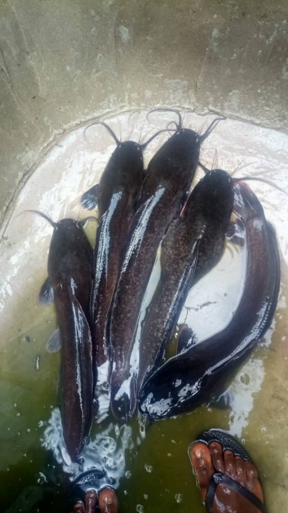
Bacterial disease of fish

FarmGrid: Fin rot

Bacterial diseases are responsible for heavy mortality in both wild and cultured fish.
Water, especially where organic loads are high, is an environment in which many bacteria could thrive.
In many fish diseases, bacteria have been found to be associated with the host, generally as secondary invaders and sometimes as primary causative agents.

Fin rot is caused by various bacteria within this group, including Cytophaga , Flexibacter, and Flavobacterium columnare.

The disease is favored often by a dirty tank, poor care, or exposure to other fish which have infectious diseases.
Fin rot affects both adults and young fish. In the early stage, the infection appears like a spreading white line on the margin of the fin, which eventually putrifies and disintegrates.

The fin rot may also cause your fish to become discolored and lethargic.

The disease may spread through contact and cause heavy damage.

Fin rot is also highly contagious and can spread to other fish within the tank. Therefore should be treated immediately it is diagnosed.

steps towards treatment

  1. Isolate the affected fish and put in a container with clean water
  2. Remove the other ones from the affected pond and keep in a separate container
  3. Drain the affected tank and wash thoroughly (with hot water) including all equipment such as nets.
  4. Once you have washed the tank thoroughly and let it air dry, fill with fresh clean water.
  5. You can slowly add the fish back into the tank, including the fish with fin rot. You may then add antibiotics to the tank water.
  6. Some farmers prefer treating the affected fish separately, but treatment must include the rest of the fish in the affected pond.

Prevention

Avoid overcrowding in the pond

Maintain a clean pond

Use high quality feed

Fascioliasis in man : zoonosis

It belongs to the group of foodborne trematode infections and is a zoonosis, meaning an animal infection that may be transmitted to humans.

The two species of trematodes that cause fascioliasis include Fasciola hepatica and F. gigantic, leaf-shaped worms, large enough to be visible to the naked eye.

Fasciola affects mainly the liver and bile duct of the host ( in this case human).

Life cycle of fasciola involves a definitive host (where the adult worm lives), an intermediate host (where the larval stages of the worm develop) and a carrier (suitable plants).

The following animals can serve as definitive hosts cattle, sheep, buffaloes, donkeys, pigs, horses, goats, camels, llamas and other herbivores. Man is also a definitive host for this parasite.

Fresh Water snails such as Lymnaea sp serve as intermediate hosts for the parasite.

When infected animals defecate in fresh-water body. The eggs of the parasite are released in the faeces and hatch into larvae that lodge in some fresh water snail which is the intermediate host.

Once inside the snail, the larva multiplies and are released into the water where they swim to suitable plants and form cysts on the leaves of such plants (and that includes vegetables).

The final hosts such as man and livestock get the parasite by eating such vegetables or plants containing the parasite (metacercaria).

Ingestion of free metacercariae floating on water may also be a possible mode of transmission or when using utensils washed with contaminated water.

People at risk include people who eat a lot of salad vegetables which are not properly washed and are undercooked

Symptoms of the disease in man

Acute phase

The immature worms penetrate the intestinal wall and the peritoneum .

From here, they penetrate the liver’s surface and eat their way through its tissues until they reach the bile ducts where they localise.

The process of getting to the liver leads to a lot of complications including damage to liver cells and internal haemorrhage or bleeding.

Notable symptoms include fever, nausea, a swollen liver, skin rashes and extreme abdominal pain.

Chronic phase

This begins when the parasite matures in the bile duct and begins to reproduce and release eggs that will start another cycle of transmission.
Symptoms include intermittent pain, jaundice and anaemia.
Patients with chronic infections experience hardening of the liver (fibrosis) as a result of the long-term inflammation.

The World Health Organization estimates that about 2.4 million people are affected by the disease and several million people are at risk

Prevention and Treatment

Promoting cultivation of vegetables in water free from faecal matters and thorough washing and cooking of vegetables before consumption.

Veterinary public health measures , including diagnosis and treatment of domestic animals.

Environmental measures such as containment of the snail intermediate hosts and drainage of grazing lands.

Maintain safe drinking water.

Treatment for man is done with WHO recommended drug triclabendazole. The drug is effective against the adult and larval stages of the parasite.

RABIES

World Rabies day 2019

The things you must know

Rabies is an infectious viral disease that is almost always fatal following the onset of clinical symptoms.
In up to 99% of cases, domestic dogs are responsible for rabies virus transmission to humans.
Yet, rabies can affect both domestic and wild animals such as foxes, bats and raccoons . It is spread to people through bites or scratches, usually via saliva of infected animals.

Rabies is a zoonotic disease. This means that rabies is one of the diseases that animals suffer from and can be transmitted to humans.

Rabies is one of the neglected tropical diseases that predominantly affects poor and vulnerable populations who live in remote rural locations.
Although effective human vaccines and immunoglobulins exist for rabies, they are not readily available or accessible to those in need. Globally, rabies deaths are rarely reported and children between the ages of 5 to 15 years are frequent victims of It.

Every year, more than 15 million people worldwide receive a post-bite vaccination. This is estimated to prevent hundreds of thousands of rabies deaths annually.

Prevention

  1. Eliminating rabies in dogs

Rabies is a vaccine-preventable disease. Vaccinating dogs is the most cost-effective strategy for preventing rabies in people.
Dog vaccination reduces deaths attributable to rabies and the need for Post Exposure prevention as a part of dog bite patient care.

FarmGrid calls on veterinarians to administer rabies vaccine at a reduced price to enable more people afford to vaccinate their dogs.

  1. Awareness on rabies and preventing dog bites

Education on dog behaviour and bite prevention for both children and adults is an essential extension of a rabies vaccination programme and can decrease both the incidence of human rabies and the financial burden of treating dog bites.

Increasing awareness of rabies prevention and control in communities should include education and information on responsible pet ownership, how to prevent dog bites, and immediate care measures after a bite.

  1. Elimination of stray dogs or being careful while getting closer to a stray dog.

Most stray dogs are carriers of this deadly virus. Human beings are therefore advised as a preventive measure, to stay away from stray dogs. Do not approach or talk to them and do not try to scare them. Just avoid them.

  1. Preventive immunization in people

Human rabies vaccines exist for pre-exposure immunization.
These are recommended for people in certain high-risk occupations such as Veterinarians, laboratory workers handling live rabies and rabies-related (lyssavirus) viruses; and people (such as animal disease control staff and wildlife rangers) whose professional or personal activities might bring them into direct contact with bats, carnivores, or other mammals that may be infected.

Pre-exposure immunization is also recommended for travellers to rabies-affected, remote areas who plan to spend a lot of time outdoors involved in activities such as caving or mountain-climbing.

Symptoms

The incubation period for rabies is typically 2–3 months but may vary from 1 week to 1 year, dependent upon factors such as the location of virus entry and viral load.
Initial symptoms of rabies include a fever with pain and unusual or unexplained tingling, pricking, or burning sensation (paraesthesia) at the wound site.
As the virus spreads to the central nervous system, progressive and fatal inflammation of the brain and spinal cord develops.
Rabies virus is neurotropic, this means that the virus affects the nerves that conduct messages to the brain and spinal cord.
Thus signs typical of CNS (brain) disturbance such as dizziness, hallucinations, muscle spasm or paralysis, mental confusion, seizures, are seen

Diagnosis

Current diagnostic tools are not suitable for detecting rabies infection before the onset of clinical disease, and unless the rabies-specific signs of hydrophobia or aerophobia are present, clinical diagnosis may be difficult. Human rabies can be confirmed intra-vitam and post mortem by various diagnostic techniques that detect whole viruses, viral antigens, or nucleic acids in infected tissues (brain, skin, urine, or saliva).

Transmission

People are usually infected following a deep bite or scratch from an animal with rabies, and transmission to humans by rabid dogs accounts for 99% of cases.
Africa and Asia have the highest rabies burden in humans and account for 95% of rabies deaths, worldwide.

Transmission can also occur when infectious material – usually saliva – comes into direct contact with human mucosa or fresh skin wounds. Human-to-human transmission through bites is theoretically possible but has never been confirmed.

NOTE; Contracting rabies through consumption of raw meat or animal-derived tissue has never been confirmed in humans. But do not attempt to be the first confirmed case.

Post-exposure prophylaxis (PEP)

Post-exposure prophylaxis (PEP) is the immediate treatment of a bite victim after rabies exposure. This prevents virus entry into the central nervous system, which results in imminent death. PEP consists of:

1) Extensive wound washing
This involves first-aid of the wound that includes immediate and thorough flushing and washing of the wound for a minimum of 15 minutes with soap and water, detergent, povidone iodine or other substances that kill the rabies virus.
2) Effective treatment of the exposed patient by trained professionals.

NOTE ; The vaccination status of the suspect animal should not be the deciding factor when considering to initiate treatment. This can be the case if dog vaccination programmes are not being sufficiently regulated or followed out of lack of resources or low priority.

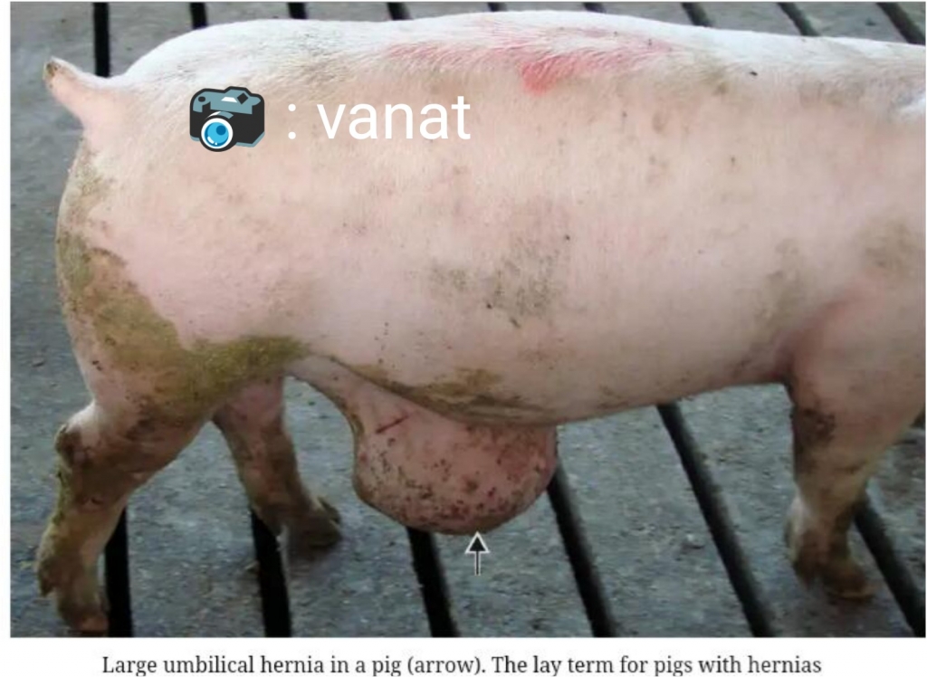
Umbilical hernia

A hernia is an abnormal protrusion of an organ or tissue through a defect or natural opening in the covering skin or muscle. Hernias often cause welfare problems as well as economic loss.

Of many congenital abnormalities, ruptures at the umbilicus or the inguinal canal are most common. They are considered to be developmental defects yet have a very low heritability

Clinical signs.
Swellings 30 – 200mm in diameter protruding from the umbilicus and abdomen, or below and in front of the testicles or in the groin (inguinal rupture).
If the swellings are large trauma to the skin may cause ulcerations particularly umbilical ruptures.

Diagnosis
1) Visual evidence of the swelling .
2) Reduction of the bowel contents in the rupture when squeezed back into the abdomen.

Causes
Umbilical hernias can sometimes be traced back to a particular boar in which case he should be culled. Environmental factors can increase the incidence of umbilical Hernias.

If there is a problem (more than 2% of pigs are affected ) consider the following:

Are prostaglandins used to synchronise farrowings. If so check that piglets are not being pulled away from the sow at farrowing and the cord stretched abnormally.

Is navel bleeding occurring on the farm? Are naval clips being used to prevent bleeding? If so make sure they are not placed close up to the skin otherwise the tissues will be damaged and weakened.
Are stocking densities high and increase abdominal pressure?
In cold weather do the pigs huddle thereby increasing abdominal pressure.
Check records to see if the boar and the sow are related.

Treatment and management
Inguinal ruptures are not as important a problem unless they become very large

If the rupture is large and the pig is on a concrete floor or slats it should be moved to a soft bedded area so that the overlying skin does not become sore and ulcerated.

Examine navels at births and two days later to see if there are any abnormalities.

Reduce stocking density of a pen if Hernias often occur in the pen.
Cull pigs with known history of hernia to avoid reoccurrence.

Veterinarians can also reduce Hernias surgically.

Get a veterinarian today from Farm Grid by requesting through our facebook page

http://facebook.com/farmgrid

Introduction

Abortion means the premature expulsion of dead or non-viable foetus, leading to foetal loss

There is often alarm when an abortion is seen but it should be remembered that there can be loss of embryos at any time during early pregnancy, which often go unseen.

Embryo loss or abortion can be considered in three main groups:

During the period from fertilisation to implantation
During the period of implantation at around 14 days post-service to 35 days.
During the period of maturation, which results in premature farrowing.
Losses can take place at any stage from approximately 14 days after mating, when implantation has taken place, through to 110 days of pregnancy.

Pregnancy is maintained through hormonal changes initiated by the implantation of embryos at day 14. These changes allow the corpus luteum (the body from which the egg is released) in the ovary to develop and produce hormones essential to maintaining pregnancy, such as progesterone. The presence of the corpus luteum is necessary to maintain the pregnancy throughout the whole of the gestation period. The loss or failure of the corpus luteum through any cause initiates the farrowing process, hence an abortion, or if near to term, a premature farrowing.

Natural biological failures of pregnancy occur across all species due to a variety of causes. In healthy sow herds, abortions observed by farmworkers occur in less than 1 out of 100 pregnancies. The cost of an abortion can be calculated quite easily, taking into account the cost of feeding the sow during a pregnancy period and the loss of margin over feed on the loss of the piglets.

Clinical signs
Occurs in breeding female adult pigs (sows and breeding gilts). Usually less than 2% of sows affected. .

Sow may show signs of illness (lethargy; inappetence; diarrhoea/scours) or may be completely normal.
Sows may show clinical signs of a specific disease, e.g. Classical swine fever or Erysipelas.
The delivery of a premature litter with or without mummified pigs.
Mucus, blood, pus discharges from the vulva.
Sow/breeding gilt is not in pig as indicated by occurrence of next heat (most fertile stage of menstrual cycle).

Diagnosis
There are three parts to the investigations that must be carried out:
1 collect information about the individual sows(Sow health history) ;
2 request post mortem examinations and serological tests;
3 assess the clinical evidence and feeding procedures in the herd.
The object of these is to identify the area of failure and by management studies, examination of records, clinical examinations, and laboratory tests, the cause may be identified.

Sow health history
It is important to study the herd history and environment. For example, is there is a seasonal effect or an association with a particular area of the housing or management practice. You should also note the clinical state of the sow at the time of abortion. Does the sow show other clinical signs or is she apparently normal?

You should examine the aborted foetuses too. Are they fresh with no signs of any decomposition, or are they decomposing or mummified? Such observations, particularly if recorded over a period, may be of help to your veterinarian in leading to a possible diagnosis of the cause of the abortion.

Post-mortem and laboratory examinations
Fresh, aborted foetuses should be submitted to a competent diagnostic laboratory where examinations can be carried out for evidence of viral and bacterial infections, together with histological examinations and toxic studies. In many cases the end results of post-mortem and serological tests do not identify any particular infectious organism, which may seem disappointing. However, it is useful in telling us what is not present.

Clinical examinations
Of all the examinations carried out, clinical observations are perhaps the most important. By using the recorded information on individual cases and collating this to the problem group of sows, it then may become possible to differentiate clinically between an infectious and a non-infectious cause.

Causes of Abortion in Pigs
Infectious causes
Infectious agents can bring about abortion in three ways:

1) They can invade the placenta, cause inflammation (placentitis) and sometimes necrosis (tissue death), cutting off the nutrient and oxygen supply to the foetus.
2) They can invade the foetus and kill it.
3) They can multiply elsewhere in the body causing fever and sometimes toxaemia (toxins in the blood).
The major infectious bacterial diseases which spread through herds and cause abortion are brucellosis and leptospirosis. These infectious agents affect increasing numbers of sows in the herd with typical clinical signs of the disease.

There is, however, a second group of bacteria which can be described as opportunist invaders which cause embryo mortality or abortion in individual sows and sporadically in small groups of sows. They do not spread through the herd like brucella or leptospira. They are often mixed infections (i.e. several different species involved). If these opportunist bacterial infections occur in sufficient numbers of sows they can become a herd problem.

They are often normal inhabitants of the vagina or the boars prepuce and their identification following pathological examinations needs careful interpretation. An emerging syndrome in this category occurs when high numbers of bacteria are deposited into the anterior vagina, particularly towards the end of the heat period, by the boar. Such bacteria include klebsiella, streptococci, staphylococci and possibly leptospira. In such cases, careful clinical examination of sows between 14 and 21 days post-mating will sometimes reveal a tacky discharge on the vulva, which may not necessarily be very obvious. Such sows should be identified, and if they are returning out of cycle, it is likely that embryo loss is taking place.

Examples of infectious diseases that can cause abortion:

Influenza virus.
Leptospira.
Specific bacteria, e.g. E. coli, klebsiella, streptococci, pseudomonas.
Parasite burdens.
Cystitis, nephritis.

Non-infectious causes
Seasonal infertility
Experiences have shown that 70% of all abortions fall into this category. Abortions, anoestrus and sows found not in pig commonly occur during the period of summer infertility when sunlight is intense and the weather is hot. Ultra violet radiation may also cause regression of the corpus luteum particularly in white breeds. This is particularly evident in outdoor sows where levels of pregnancy failure may reach 15-30%. In such cases the abortions are so early that the foetuses are either not seen or there is progressive embryo mortality and a delayed return to oestrus. This is well recognised with summer infertility and the autumn abortion syndrome, where environmental factors are likely to cause the corpus luteum to fail.

2) Decreasing daylight length, poor lighting
To maintain a viable pregnancy requires constant daylight length. Ideally this should be 12-16 hours per day. Light intensity experienced by the sow can be affected by a number of environmental inadequacies, for example, poor lighting in the first place, followed by fly faeces and dust on lamps gradually reducing the availability of light. High walls surrounding animals, or automatic feeders in front of sows produce shadows. A simple tip here is to make sure that you can read a newspaper in the darkest parts of the building at sow eye level. If not, then problems may start. Painting the roofs and walls white to increase the reflection of light is one way of improving the environment and on a number of occasions abortions have ceased after such simple improvements.

3) Low temperatures, chilling, draughts
Wet, damp environments or high air movement cause chilling and increase demands for energy. An important feature of environmental abortions is that the sow remains normal, often eating feed in the morning, and expelling the litter in the afternoon.

4) Poor nutrition
If the metabolism of sows are allowed to progress to a negative energy or catabolic state so that they are having to use their body tissues to maintain the energy equilibrium, then individual susceptible animals may abort. Clinical examinations will identify possible changes in the environment. For example, the removal of bedding, poor quality feeds, or a drop in feed intake. The latter may simply be associated with a change in stockworkers. Outbreaks of abortion may occur when there are changes from pellet feeding to meal feeding, or where feed is presented by volume and not by weight. The aborted foetuses are perfectly normal and the sow shows no signs of illness. The underlying initiating mechanism is regression of the corpus luteum.

4) Mouldy feeds
Toxins from mouldy feed can cause abortions. The fungus can be at a low level in one of the feed constituents and barely detectable by visual examination and yet produce powerful toxins. These when ingested have toxic effects which may cause abortion. This form of fungal poisoning is called mycotoxicosis. The fungus may also be readily seen growing as a mould in feed lines, bags of feed or wet feed systems and their distributing vessels and pipes.

5) Stress
Bullying and fighting are often forgotten as predisposing factors of stress in individual sows.

6) No boar contact
The presence of the boar and his pheromones (male chemical hormones) . Pheromones are required to maintain pregnancy in individual susceptible disadvantaged sows.

7) Lameness
Lameness and pain, particularly from abscesses in the feet or leg weakness (osteochondrosis) can also cause the corpus luteum to regress due to stress.

Treatment and management
Records help to identify reproductive problems. These should include information on:

Age (or parity) profile of the herd.
Sow number.
Feed and amounts given.
Clinical observations of the sow and any disease history.
Boar used during mating; date of service.
Failure to come on heat.
Date of abortion and condition of aborted piglets.
Culling rates.
Bleeding and discharges from the vulva.
Lameness.
Litter sizes.
Mastitis, lack of milk, swollen udders.
Deaths and their likely causes.
Poor conformation.

Feed
Increase feed intake from days 3 to 21 after mating.

Heat and light management
The outdoor breeding female should always be derived from at least one pigmented parent.
Provide extensive shades so that the sows can protect themselves from the sun.
Site the arks in the wind direction so that with open ends cooling can take place.
Provide extensive well-maintained wallows suitably sited so that sows do not have too far to reach them.

Contact with the boar
Boar presence in the dry sow accommodation is recommended from the day of service through to the day of farrowing. The boar should be mixed in or have access to the group for at least the first 21 days of pregnancy. There is clear evidence that this will improve farrowing rates particularly if they are associated with summer infertility. If sows are individually housed the boar should be allowed to move down the passages and make social contact daily.
Increase the mating programme by 10-15% over the anticipated period of infertility.
Because boar semen can be affected follow each natural mating 24 hours later by purchased AI.

Mycotoxin management
Are the bags of feed kept in a dry cool or wet warm place?
Are you ever tempted to give feed to sows that has been slightly moody?

Most importantly, contact your Veterinarian anytime a gilt or sow abort

Human echinococcosis is a parasitic disease caused by tapeworms of the genus Echinococcus.
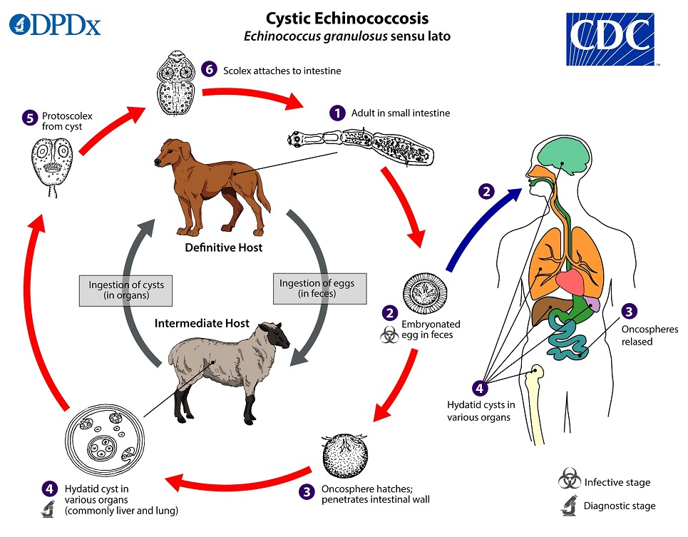
The lifecycle of E. granulosus involves dogs and wild carnivores as a definitive host for the adult tapeworm. Definitive hosts are where parasites reach maturity and reproduce.
The two most important forms of the disease in humans are cystic echinococcosis (hydatidosis) caused by Echinococcus granulosus and alveolar echinococcosis caused by E. multilocularis.
Echinococcosis is expensive and often complicated to treat, and may require extensive surgery and/or prolonged drug therapy.
Communities that practice sheep farming experience the highest risk to humans being infected. however, cattle, horses, pigs, goats, and camels are also potential intermediate hosts.
Humans are infected through ingestion of parasite eggs in contaminated food, water or soil, or after direct contact with animal hosts such as dogs and wild carnivores.
Transmission
Herbivores and some omnivores act as intermediate hosts and become infected by ingesting the parasite eggs in contaminated food and water, and the parasite then develops into larval stages in the viscera.
Carnivores such as dogs are the final hosts and are infected through the consumption of viscera of intermediate hosts that contain the parasite larvae.
Humans get infected same way as the herbivores and omnivores.
Signs and symptoms of hydatid disease in man
The rate at which symptoms appear typically depends on the location of the cyst
Human infection with E. granulosus leads to the development of one or more hydatid cysts in one or several organs such as the liver and lungs, the bones, kidneys, spleen, muscles and the brain.
Signs depends on the location of the hydatid cyst.

Abdominal pain and vomiting are commonly seen when hydatids occur in the liver.
If the lung is affected, clinical signs include chronic cough, chest pain and shortness of breath.
Rupture of the cysts can potentially lead to anaphylactic shock or cyst dissemination to various organs.
Echinococcus multilocularis affects the liver as a slow growing, destructive tumor, often with abdominal pain and biliary obstruction being the only manifestations evident in early infection.
Many rodents may serve as intermediate hosts for E. multilocularis.
Treatment
According to the WHO
There are 4 options for the treatment of cystic echinococcosis:
1. percutaneous treatment of the hydatid cysts with the PAIR (Puncture, Aspiration, Injection, Re-aspiration) technique;
2. surgery
3. anti-infective drug treatment
4. “watch and wait”.
Prevention
Improper disposal of carcasses and offal after home slaughter makes dogs readily have access to offal from livestock, thus completing the parasite cycle of Echinococcus granulosus and putting communities at risk of cystic echinococcosis
Periodic deworming of dogs with praziquantel (at least 4 times per year), improved hygiene in the slaughtering of livestock (including the proper destruction of infected offal), and public education campaigns remains the best prevention option.

Introduction
Large volumes of gas are generated in the rumen on regular basis by microbial fermentation .
Bloat occurs when bulk of this gas is not eliminated from the rumen by eructation or belching which is usually the means.
The rumen and reticulum becomes abnormally distended or enlarged with gases derived from rumen fermentation.
Frothy bloat may result in animals on pasture containing high amounts of alfalfa or clover or similar legumes which are rapidly digested in the rumen producing large bubbles of gas.
Frothy bloat can also occur in animals fed high levels of grains and concentrate rations.
Frothy bloat occurs after rapid ingestion of high protein, highly digestible feed that results in formation of excess amounts of stable foam in the rumen.
Free gas bloat occurs when the animal is unable to eructate free gas in the rumen.
The condition maybe due to partial obstruction of the esophagus by foreign bodies or tumors.
Consequences of bloat
Distension of the rumen puts pressure on thoracic and abdominal organs reducing blood flow to such organs.
Also, pressure on the diaphragm interferes with lung function leading to death due to hypoxia ( tissues and organs deprived of adequate oxygen).
Clinical signs
Left side abdominal distension
Affected animal stops eating and shows signs of distress
Breathing difficulties (rapid breathing)
Reluctance to move and recumbency and death
Cattle with tetanus, rabies, oesophageal obstruction and ruminal acidosis may also develop bloat.
With rabies there is a behaviour issue, with oesophageal obstruction there is profuse salivation and with acidosis there is also dehydration and liquid brown faeces.
In tetanus, the animal appears stiff.

Treatment
Bloat is usually a life-threatening situation in livestock. A veterinarian should be sought immediately.
Insertion of a rumen trochar through the left flank into rumen is sometimes done but is often followed by complications such as peritonitis.
In less severe cases, stomach tube could be passed down the oesophagus into the rumen to relieve it of free gas.
Antifoaming agents such as vegetable oils, paraffin, etc can be delivered directly into the rumen via stomach tube or by drenching in cases of frothy bloat.
A veterinarian may recommend rumenotomy if none of the above worked.
The best control is to maintain balance between hay and legume pastures. Usually it is better to feed hays before introducing the animals to pasture.
Another way is to incorporate antifoaming agents in their feed or small pasture to prevent bloat.
Grains given to feedlot animals should not be finely ground as this induces bloat. Ensure to include roughages such as straw or hay in feedlot rations
As much as possible, minimize competition during feeding
Cattle areas must be kept free of wastes such as plastic, string, rope and wire that may lodge somewhere in the tract and cause bloat through obstruction.