Human echinococcosis is a parasitic disease caused by tapeworms of the genus Echinococcus.
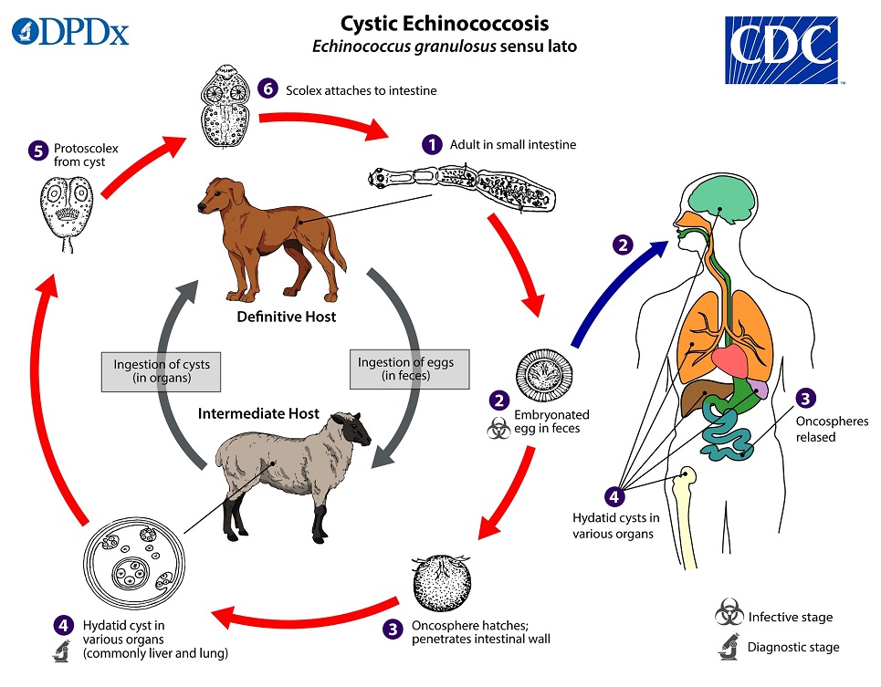
The lifecycle of E. granulosus involves dogs and wild carnivores as a definitive host for the adult tapeworm. Definitive hosts are where parasites reach maturity and reproduce.
The two most important forms of the disease in humans are cystic echinococcosis (hydatidosis) caused by Echinococcus granulosus and alveolar echinococcosis caused by E. multilocularis.
Echinococcosis is expensive and often complicated to treat, and may require extensive surgery and/or prolonged drug therapy.
Communities that practice sheep farming experience the highest risk to humans being infected. however, cattle, horses, pigs, goats, and camels are also potential intermediate hosts.
Humans are infected through ingestion of parasite eggs in contaminated food, water or soil, or after direct contact with animal hosts such as dogs and wild carnivores.
Transmission
Herbivores and some omnivores act as intermediate hosts and become infected by ingesting the parasite eggs in contaminated food and water, and the parasite then develops into larval stages in the viscera.
Carnivores such as dogs are the final hosts and are infected through the consumption of viscera of intermediate hosts that contain the parasite larvae.
Humans get infected same way as the herbivores and omnivores.
Signs and symptoms of hydatid disease in man
The rate at which symptoms appear typically depends on the location of the cyst
Human infection with E. granulosus leads to the development of one or more hydatid cysts in one or several organs such as the liver and lungs, the bones, kidneys, spleen, muscles and the brain.
Signs depends on the location of the hydatid cyst.
Abdominal pain and vomiting are commonly seen when hydatids occur in the liver.
If the lung is affected, clinical signs include chronic cough, chest pain and shortness of breath.
Rupture of the cysts can potentially lead to anaphylactic shock or cyst dissemination to various organs.
Echinococcus multilocularis affects the liver as a slow growing, destructive tumor, often with abdominal pain and biliary obstruction being the only manifestations evident in early infection.
Many rodents may serve as intermediate hosts for E. multilocularis.
Treatment
According to the WHO
There are 4 options for the treatment of cystic echinococcosis:
1. percutaneous treatment of the hydatid cysts with the PAIR (Puncture, Aspiration, Injection, Re-aspiration) technique;
2. surgery
3. anti-infective drug treatment
4. “watch and wait”.
Prevention
Improper disposal of carcasses and offal after home slaughter makes dogs readily have access to offal from livestock, thus completing the parasite cycle of Echinococcus granulosus and putting communities at risk of cystic echinococcosis
Periodic deworming of dogs with praziquantel (at least 4 times per year), improved hygiene in the slaughtering of livestock (including the proper destruction of infected offal), and public education campaigns remains the best prevention option.
